| Citation: | Krittirash Yorseng, Suchart Siengchin, Basa Ashok, Anumakonda Varada Rajulu. Nanocomposite Egg Shell Powder with in situ Generated Silver Nanoparticles Using Inherent Collagen as Reducing Agent[J]. Journal of Bioresources and Bioproducts, 2020, 5(2): 101-107. doi: 10.1016/j.jobab.2020.04.003

|
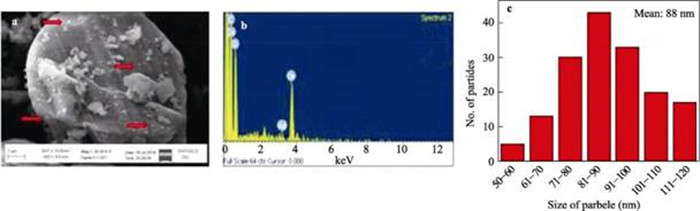
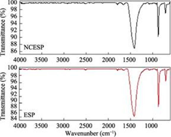
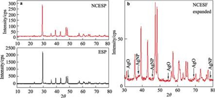
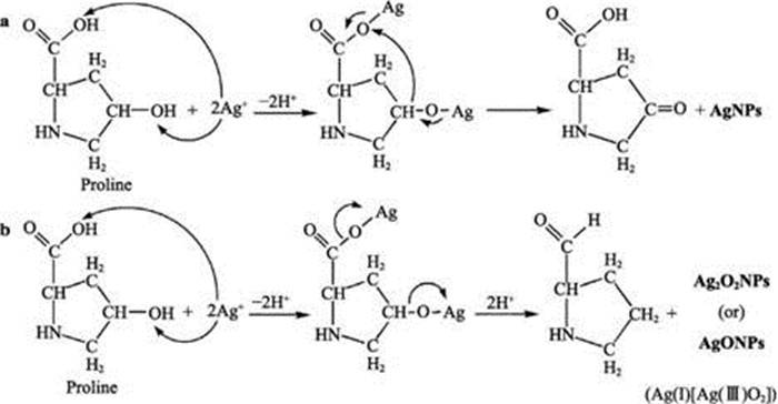
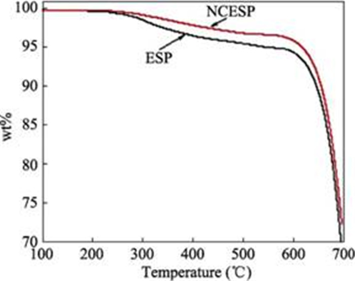
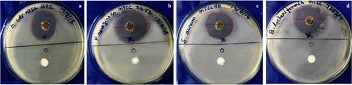
Silver nanoparticles (AgNPs) were in situ generated in poultry hen egg shell powder (ESP) by one step thermal assisted method using the inherently present collagen as a reducing agent. The nanocomposite egg shell powder (NCESP) with in situ generated silver nanoparticles was characterized by scanning electron microscopy (SEM), Fourier transform infrared (FT-IR) spectroscopy, X-ray diffraction (XRD), thermogravimetric analysis (TGA) and antibacterial tests. The prepared NCESP had the spherical AgNPs in the size range of 50Ƀ120 nm with most of them from 81 nm to 90 nm. Further, the average size of the AgNPs generated in the NCESP was 88 nm. The X-ray analysis indicated the presence of both AgNPs and AgO nanoparticles (AgONPs) in the NCESP. The possible mechanism of generation of AgNPs and AgONPs in the NCESP was also proposed. The thermal stability of the NCESP was found to be higher than that of the ESP. The NCESP exhibited excellent antibacterial activity against both the Gram negative and positive bacteria. The NCESP made from poultry waste ESP can be utilized as a low-cost antibacterial cleaning powder for house ware and also as low-cost antibacterial filler in polymer matrices to make antibacterial hybrid nanocomposites.

|
Ahmed, S., Saifullah, Ahmad, M., Swami, B.L., Ikram, S., 2016. Green synthesis of silver nanoparticles using Azadirachta indica aqueous leaf extract. J. Radiat. Res. Appl. Sci. 9, 1-7. doi: 10.1016/j.jrras.2015.06.006
|
|
Asadi, S., Charati, F.R., Akbari, R., Razavi, S.A., 2018. Green synthesis of silver nanoparticles using Taxus baccata Leaves extract and identify its specifications. J. Mater. Environ. Sci. 9, 2798-2803.
|
|
Ashok, B., Naresh, S., Reddy, K.O., Madhukar, K., Cai, J., Zhang, L., Rajulu, A.V., 2014. Tensile and thermal properties of poly(lactic acid)/eggshell powder composite films. Int. J. Polym. Anal. Charact. 19, 245-255. doi: 10.1080/1023666X.2014.879633
|
|
Ashok, B., Obi Reddy, K., Yorseng, K., Rajini, N., Hariram, N., Siengchin, S., Varada Rajulu, A., 2018. Modification of natural fibers from Thespesia lampas plant by in situ generation of silver nanoparticles in single-step hydrothermal method. Int. J. Polym. Anal. Charact. 23, 509-516. doi: 10.1080/1023666X.2018.1486270
|
|
Banerjee, P., Satapathy, M., Mukhopahayay, A., Das, P., 2014. Leaf extract mediated green synthesis of silver nanoparticles from widely available Indian plants:synthesis, characterization, antimicrobial property and toxicity analysis. Bioresour. Bioprocess. 1, 3. doi: 10.1186/s40643-014-0003-y
|
|
Behravan, M., Hossein Panahi, A., Naghizadeh, A., Ziaee, M., Mahdavi, R., Mirzapour, A., 2019. Facile green synthesis of silver nanoparticles using Berberis vulgaris leaf and root aqueous extract and its antibacterial activity. Int. J. Biol. Macromol. 124, 148-154. doi: 10.1016/j.ijbiomac.2018.11.101
|
|
Belbachir, K., Noreen, R., Gouspillou, G., Petibois, C., 2009. Collagen types analysis and differentiation by FTIR spectroscopy. Anal. Bioanal. Chem. 395, 829-837. doi: 10.1007/s00216-009-3019-y
|
|
Birusanti, A.B., Mallavarapu, U., Nayakanti, D., Espenti, C.S., Mala, S., 2019. Sustainable green synthesis of silver nanoparticles by using Rangoon creeper leaves extract and their spectral analysis and anti-bacterial studies. IET Nanobiotechnology 13, 71-76. doi: 10.1049/iet-nbt.2018.5117
|
|
Camacho, N.P., West, P., Torzilli, P.A., Mendelsohn, R., 2001. FTIR microscopic imaging of collagen and proteoglycan in bovine cartilage. Biopolymers 62, 1-8. doi: 10.1002/1097-0282(2001)62:1<1::AID-BIP10>3.0.CO;2-O
|
|
Chen, B., Yan, L., Liu, X., Worral, J.L., 2016. Poultry keratin based decolorants for dyeing waste water treatment. J. Bioresour. Bioprod. 1, 30-35.
|
|
Chen, Y., Cao, X.D., Chang, P.R., Huneault, M.A., 2008. Comparative study on the films of poly(vinyl alcohol)/pea starch nanocrystals and poly(vinyl alcohol)/native pea starch. Carbohydr. Polym. 73, 8-17. doi: 10.1016/j.carbpol.2007.10.015
|
|
Feng, Y., Ashok, B., Madhukar, K., Zhang, J.M., Zhang, J., Reddy, K.O., Rajulu, A.V., 2014. Preparation and characterization of polypropylene carbonate bio-filler (eggshell powder) composite films. Int. J. Polym. Anal. Charact. 19, 637-647. doi: 10.1080/1023666X.2014.953747
|
|
Galván-Ruiz, M., Hernández, J., Baños, L., Noriega-Montes, J., Rodríguez-García, M.E., 2009. Characterization of calcium carbonate, calcium oxide, and calcium hydroxide as starting point to the improvement of lime for their use in construction. J. Mater. Civ. Eng. 21, 694-698. doi: 10.1061/(ASCE)0899-1561(2009)21:11(694)
|
|
Haroon, H.I., Elbadawi, A.A., Siddig, M.A., Abuelhassan, H.H., Sabah Elkhair, M.K., 2015. Studying the physical characters of eggshell and recycling hen's egg waste as powder for cleaning used in household wares. Nova J. Med. Biol. Sci. 4, 1-10.
|
|
Ibrahim, H.M.M., 2015. Green synthesis and characterization of silver nanoparticles using banana peel extract and their antimicrobial activity against representative microorganisms. J. Radiat. Res. Appl. Sci. 8, 265-275. doi: 10.1016/j.jrras.2015.01.007
|
|
Iram, J., 2019. FTIR analysis of egg shell of pigeon Columba livia. Int. J. Res. Appl. Sci. Eng. Technol. 7, 1595-1596. https://www.ijraset.com/fileserve.php?FID=20631
|
|
Jawaid, M., Siengchin, S., 2019. Hybrid composites:a versatile materials for future. Applied Science and Engineering Progress 12, 223. http://d.old.wanfangdata.com.cn/Periodical/nmyj-z201808002
|
|
Karunagaran, V., Rajendran, K., Sen, S., 2014. Antimicrobial activity of biosynthesized silver oxide nanoparticles. Journal of Pure and Applied Microbiology 4, 3263-3268. http://www.wanfangdata.com.cn/details/detail.do?_type=perio&id=ff3cd7f24e1f7a628e703d791f4aa3bf
|
|
Kishanji, M., Mamatha, G., Obi Reddy, K., Varada Rajulu, A., Madhukar, K., 2017. In situ generation of silver nanoparticles in cellulose matrix using Azadirachta indica leaf extract as a reducing agent. Int. J. Polym. Anal. Charact. 22, 734-740. doi: 10.1080/1023666X.2017.1369612
|
|
Lin, X., Wang, J., Han, X., Wu, M., Kuga, S., Huang, Y., 2017. Use of lignin and hemicelluloses for facia synthesis of gold, platinum and palladium nanoparticles. J. Bioresour. Bioprod. 2, 149-152.
|
|
Ly, N., Seo, C., Joo, S.W., 2016. Detection of copper(Ⅱ) ions using glycine on hydrazine-adsorbed gold nanoparticles via Raman spectroscopy. Sensors 16, 1785. doi: 10.3390/s16111785
|
|
Makvandi, P., Nikfarjam, N., Sanjani, N.S., Qazvini, N.T., 2015. Effect of silver nanoparticle on the properties of poly(methyl methacrylate) nanocomposite network made by in situ photoiniferter-mediated photopolymerization. Bull. Mater. Sci. 38, 1625-1631. doi: 10.1007/s12034-015-0959-z
|
|
Meejoo, S., Maneeprakorn, W., Winotai, P, 2006. Phase and thermal stability of nanocrystalline hydroxyapatite prepared via microwave heating. Thermochimica Acta 447, 115-120. doi: 10.1016/j.tca.2006.04.013
|
|
Muthulakshmi, L., Rajini, N., Nellaiah, H., Kathiresan, T., Jawaid, M., Varada Rajulu, A., 2017. Experimental investigation of cellulose/silver nanocomposites using in situ generation method. J. Polym. Environ. 25, 1021-1032. doi: 10.1007/s10924-016-0871-7
|
|
Ok, Y.S., Lee, S.S., Jeon, W.T., Oh, S.E., Usman, A.R.A., Moon, D.H., 2011. Application of eggshell waste for the immobilization of cadmium and lead in a contaminated soil. Environ. Geochem. Heal. 33, 31-39. doi: 10.1007/s10653-010-9362-2
|
|
Pan, Y., Farmahini-Farahani, M., Hearn, O.P., Xiao, H., Ocampo, H., 2016. An overview of biobased polymers for packaging materias. J. Bioresour. Bioprod. 1, 106-113.
|
|
Pusphalatha, R., Ashok, B., Hariram, N., Rajulu, A.V., 2019. Nanocomposite polyester fabrics with in situ generated silver nanoparticles using tamarind leaf extract reducing agent. Int. J. Polym. Anal. Charact. 24, 524-532. doi: 10.1080/1023666X.2019.1614265
|
|
Rajesh Kumar, T.V., Murthy, J.S.R., Narayana Rao, M., Bhargava, Y., 2016. Evaluation of silver nanoparticles synthetic potential of Couroupita guianensis Aubl., flower buds extract and their synergistic antibacterial activity. 3 Biotech 6, 92. https://www.ncbi.nlm.nih.gov/pmc/articles/PMC4801843/
|
|
Sadanand, V., Rajini, N., Varada Rajulu, A., Satyanarayana, B., 2018. Effect of sunlight on the preparation and properties of cellulose/silver nanoparticle composite films by in situ method using Ocimum sanctum leaf extract as a reducing agent. Int. J. Polym. Anal. Charact. 23, 313-320. doi: 10.1080/1023666X.2018.1440915
|
|
Sadanand, V., Tian, H.F., Rajulu, A.V., Satyanarayana, B., 2017. Antibacterial cotton fabric with in situ generated silver nanoparticles by one-step hydrothermal method. Int. J. Polym. Anal. Charact. 22, 275-279. doi: 10.1080/1023666X.2017.1287828
|
|
Singhal, G., Bhavesh, R., Kasariya, K., Sharma, A.R., Singh, R.P., 2011. Biosynthesis of silver nanoparticles using Ocimum sanctum (Tulsi) leaf extract and screening its antimicrobial activity. J. Nanoparticle Res. 13, 2981-2988. doi: 10.1007/s11051-010-0193-y
|
|
Sivaranjana, P., Nagarajan, E.R., Rajini, N., Jawaid, M., Rajulu, A.V., 2017. Cellulose nanocomposite films with in situ generated silver nanoparticles using Cassia alata leaf extract as a reducing agent. Int. J. Biol. Macromol. 99, 223-232. doi: 10.1016/j.ijbiomac.2017.02.070
|
|
Sriram, T., Pandidurai, V., 2014. Synthesis of silver nanoparticles from leaf extract of Psidium guajava and its antibacterial activity against pathogens. International Journal of Current Microbiology Applied Science 3, 146-152. http://www.sciencedirect.com/science/article/pii/S1995764514601711
|
|
Wang, C.Y., Xiao, P., Zhao, J.Z., Zhao, X., Liu, Y.H., Wang, Z.C., 2006. Biomimetic synthesis of hydrophobic calcium carbonate nanoparticles via a carbonation route. Powder Technol. 170, 31-35. doi: 10.1016/j.powtec.2006.08.016
|

Figures(7) / Tables(1)

Copyright © 2019 Editorial Office of Journal of Bioresources and Bioproducts
Supported by: Beijing Renhe Information Technology Co. Ltd support: info@rhhz.net


 DownLoad: